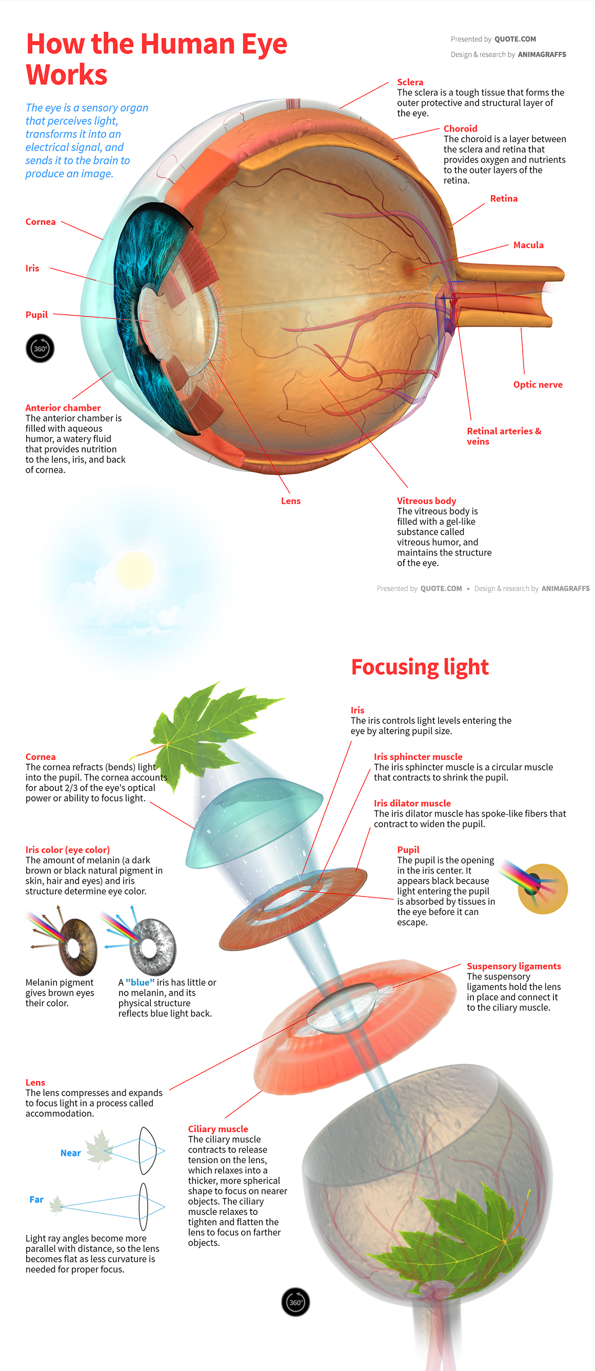
How the Human Eye Works
The eye is a sensory organ that perceives light, transforms it into an electrical signal, and sends it to the brain to produce an image.
Sclera
The sclera is a tough tissue that forms the outer protective and structural layer of the eye.
Choroid
The choroid is a layer between the sclera and retina that provides oxygen and nutrients to the outer layers of the retina.
Retina
Macula
Optic nerve
Retinal arteries & veins
Vitreous body
The vitreous body is filled with a gel-like substance called vitreous humor, and maintains the structure of the eye.
Lens
Anterior chamber
The anterior chamber is filled with aqueous humor, a watery fluid that provides nutrition to the lens, iris, and back of cornea.
Pupil
Iris
Cornea
Focusing light
Iris
The iris controls light levels entering the eye by altering pupil size.
Iris sphincter muscle
The iris sphincter muscle is a circular muscle that contracts to shrink the pupil.
Iris dilator muscle
The iris dilator muscle has spoke-like fibers that contract to widen the pupil.
Pupil
The pupil is the opening in the iris center. It appears black because light entering the pupil is absorbed by tissues in the eye before it can escape.
Suspensory ligaments
The suspensory ligaments hold the lens in place and connect it to the ciliary muscle.
Ciliary muscle
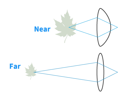
The ciliary muscle contracts to release tension on the lens, which relaxes into a thicker, more spherical shape to focus on nearer objects. The ciliary muscle relaxes to tighten and flatten the lens to focus on farther objects.
Lens
The lens compresses and expands to focus light in a process called accommodation.
Light ray angles become more parallel with distance, so the lens becomes flat as less curvature is needed for proper focus.
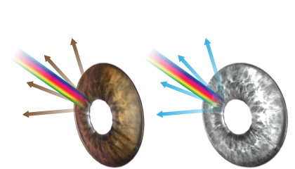
Iris color (eye color)
The amount of melanin (a dark brown or black natural pigment in skin, hair and eyes) and iris structure determine eye color.
Melanin pigment gives brown eyes their color.
A "blue" iris has little or no melanin, and its physical structure reflects blue light back.
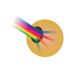
Cornea
The cornea refracts (bends) light into the pupil. The cornea accounts for about 2/3 of the eye's optical power or ability to focus light.
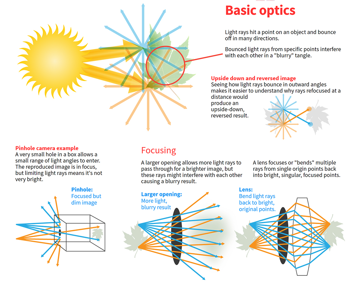
Basic optics
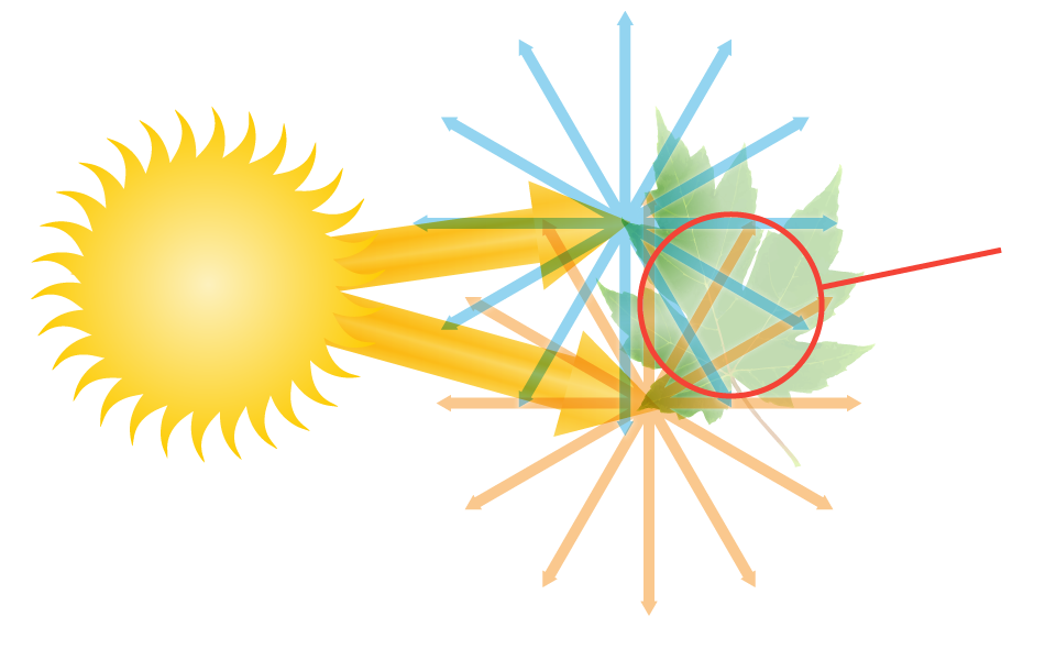
Light rays hit a point on an object and bounce off in many directions.
Bounced light rays from specific points interfere with each other in a "blurry" tangle.
Upside down and reversed image
Seeing how light rays bounce in outward angles makes it easier to understand why rays
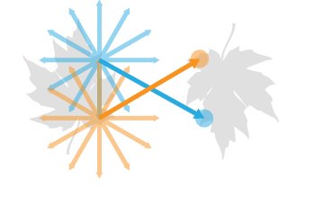 refocused at a distance would produce an upside-down, reversed result.
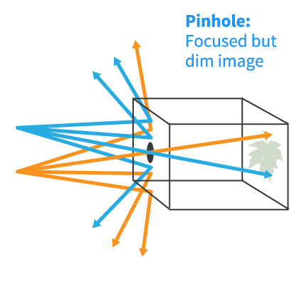
Pinhole camera example
A very small hole in a box allows a small range of light angles to enter. The reproduced image is in focus, but limiting light rays means it's not very bright.
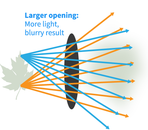
Focusing
A larger opening allows more light rays to pass through for a brighter image, but these rays might interfere with each other causing a blurry result.
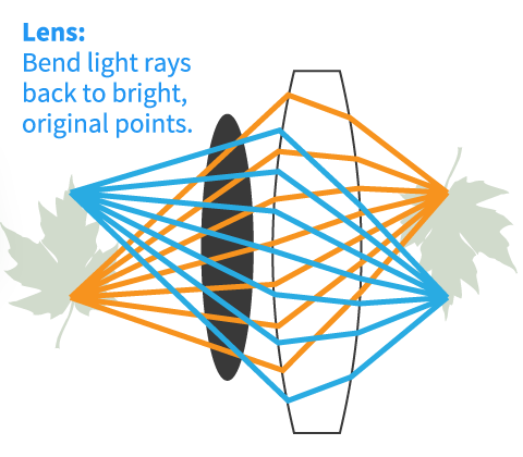
A lens focuses or "bends" multiple rays from single origin points back into bright, singular, focused points.
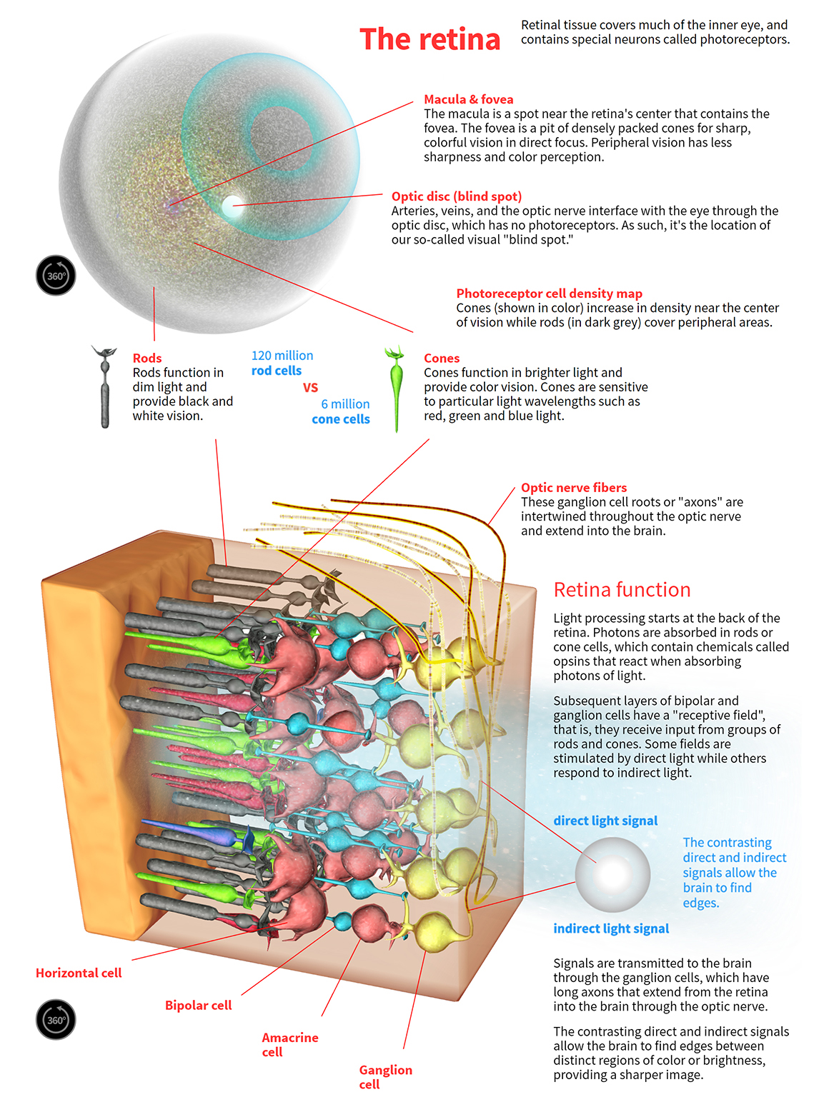
The retina
Retinal tissue covers much of the inner eye, and contains special neurons called photoreceptors.
Macula & fovea
The macula is a spot near the retina's center that contains the fovea. The fovea is a pit of densely packed cones for sharp, colorful vision in direct focus. Peripheral vision has less sharpness and color perception.
Optic disc (blind spot)
Arteries, veins, and the optic nerve interface with the eye through the optic disc, which has no photoreceptors. As such, it's the location of our so-called visual "blind spot."
Photoreceptor cell density map
Cones (shown in color) increase in density near the center of vision while rods (in dark grey) cover peripheral areas.
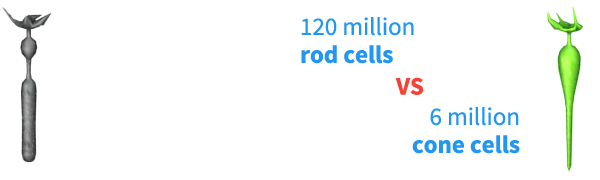
Rods
Rods function in dim light and provide black and white vision.
Cones
Cones function in brighter light and provide color vision. Cones are sensitive to particular light wavelengths such as red, green and blue light.
Optic nerve fibers
These ganglion cell roots or "axons" are intertwined throughout the optic nerve and extend into the brain.
Retina function
Light processing starts at the back of the retina. Photons are absorbed in rods or cone cells, which contain chemicals called opsins that react when absorbing photons of light.
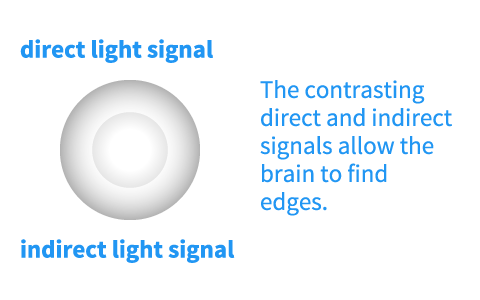
Subsequent layers of bipolar and ganglion cells have a "receptive field", that is, they receive input from groups of rods and cones. Some fields are stimulated by direct light while others respond to indirect light.
Signals are transmitted to the brain through the ganglion cells, which have long axons that extend from the retina into the brain through the optic nerve.
The contrasting direct and indirect signals allow the brain to find edges between distinct regions of color or brightness, providing a sharper image.
Ganglion cell
Amacrine cell
Bipolar cell
Horizontal cell
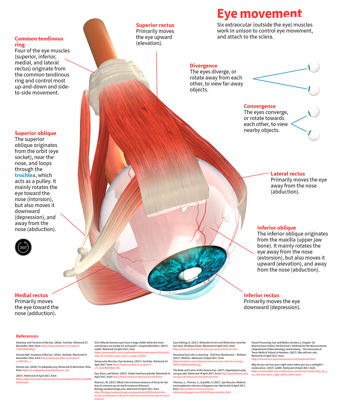
Eye movement
Six extraocular (outside the eye) muscles work in unison to control eye movement, and attach to the sclera.
Superior rectus
Primarily moves the eye upward (elevation).
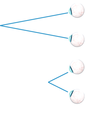
Divergence
The eyes diverge, or rotate away from each other, to view far-away objects.
Convergence
The eyes converge, or rotate towards each other, to view nearby objects.
Lateral rectus
Primarily moves the eye away from the nose (abduction).
Inferior oblique
The inferior oblique originates from the maxilla (upper jaw bone). It mainly rotates the eye away from the nose (extorsion), but also moves it upward (elevation), and away from the nose (abduction).
Inferior rectus
Primarily moves the eye downward (depression).
Medial rectus
Primarily moves the eye toward the nose (adduction).
Superior oblique
The superior oblique originates from the orbit (eye socket), near the nose, and loops through the trochlea, which acts as a pulley. It mainly rotates the eye toward the nose (intorsion), but also moves it downward (depression), and away from the nose (abduction).
Common tendinous ring
Four of the eye muscles (superior, inferior, medial, and lateral rectus) originate from the common tendinous ring and control most up-and-down and side-to-side movement.
References
- Anatomy and Function of the Eye. (2016). YouTube. Retrieved 23 November 2016, from https://www.youtube.com/watch?v=RE1MvRmWg7I
- Human A&P: Anatomy of the Eye. (2016). YouTube. Retrieved 23 November 2016, from https://www.youtube.com/watch?v=TXFt1Ikl__I
- Human eye. (2016). En.wikipedia.org. Retrieved 23 November 2016, from https://en.wikipedia.org/wiki/Human_eye
- (2017). Retrieved 14 April 2017, from http://webvision.med.utah.edu/wp-content/uploads/2011/01/2003-01Kolb.pdf
- ELI5: Why do humans eyes have a large visible white but most animal eyes are mostly iris and pupil? • r/explainlikeimfive. (2017). reddit. Retrieved 14 April 2017, from https://www.reddit.com/r/explainlikeimfive/comments/23iink/eli5_why_do_humans_eyes_have_a_large_visible/
- Extraocular Muscles | Eye Anatomy. (2017). YouTube. Retrieved 14 April 2017, from https://www.youtube.com/watch?v=f_rb6FMVHPk
- Eye, Brain, and Vision. (2017). Hubel.med.harvard.edu. Retrieved 14 April 2017, from http://hubel.med.harvard.edu/book/b8.htm
- flatness?, W. (2017). What's the minimum distance of focus for the lens of a human eye to reach maximum flatness?. Biology.stackexchange.com. Retrieved 14 April 2017, from http://biology.stackexchange.com/questions/15981/whats-the-minimum-distance-of-focus-for-the-lens-of-a-human-eye-to-reach-maximu
- Gary Heiting, O. (2017). Refractive Errors and Refraction: How the Eye Sees. All About Vision. Retrieved 14 April 2017, from http://www.allaboutvision.com/eye-exam/refraction.htm
- Structural Eye Color is Amazing – Paul Van Slembrouck – Medium. (2017). Medium. Retrieved 14 April 2017, from https://medium.com/@ptvan/structural-eye-color-is-amazing-24f47723bf9a#.b8zfrcryg
- The Rods and Cones of the Human Eye. (2017). Hyperphysics.phy-astr.gsu.edu. Retrieved 14 April 2017, from http://hyperphysics.phy-astr.gsu.edu/hbase/vision/rodcone.html
- Thomas, S., Thomas, S., & profile, V. (2017). Eye Muscles. Medical-transcriptionist-reference.blogspot.com. Retrieved 14 April 2017, from http://medical-transcriptionist-reference.blogspot.com/2012/05/eye-muscles.html
- Visual Processing: Eye and Retina (Section 2, Chapter 14) Neuroscience Online: An Electronic Textbook for the Neurosciences | Department of Neurobiology and Anatomy - The University of Texas Medical School at Houston. (2017). Nba.uth.tmc.edu. Retrieved 14 April 2017, from http://nba.uth.tmc.edu/neuroscience/s2/chapter14.html
- Why do you not lose your night vision when you use a red light? • r/askscience. (2017). reddit. Retrieved 14 April 2017, from https://www.reddit.com/r/askscience/comments/1u6sf5/why_do_you_not_lose_your_night_vision_when_you/
Share / embed code
• Copy/paste the code below to share this project on your site (in an iframe).
• We only require a link back to this page and name attribution (ex: "by Quote.com")
Sharing images
(click for large versions)